
When There Is No Air
What happens to cancer cells?

“Your cancer is back.” I am assuming that is what this patient was told.
In March 2022, a 62-year-old man presented with stage 2 stomach cancer at a Chinese hospital. Doctors performed surgery to remove the lower half of his stomach and the nearby lymph nodes, then followed up with six cycles of chemotherapy. Sadly, by December 2023, the cancer had returned. He received four cycles of systemic immunochemotherapy (CAPOX + sintilimab). By March 2024, his scans revealed stable disease, meaning that the cancer was neither growing nor shrinking.
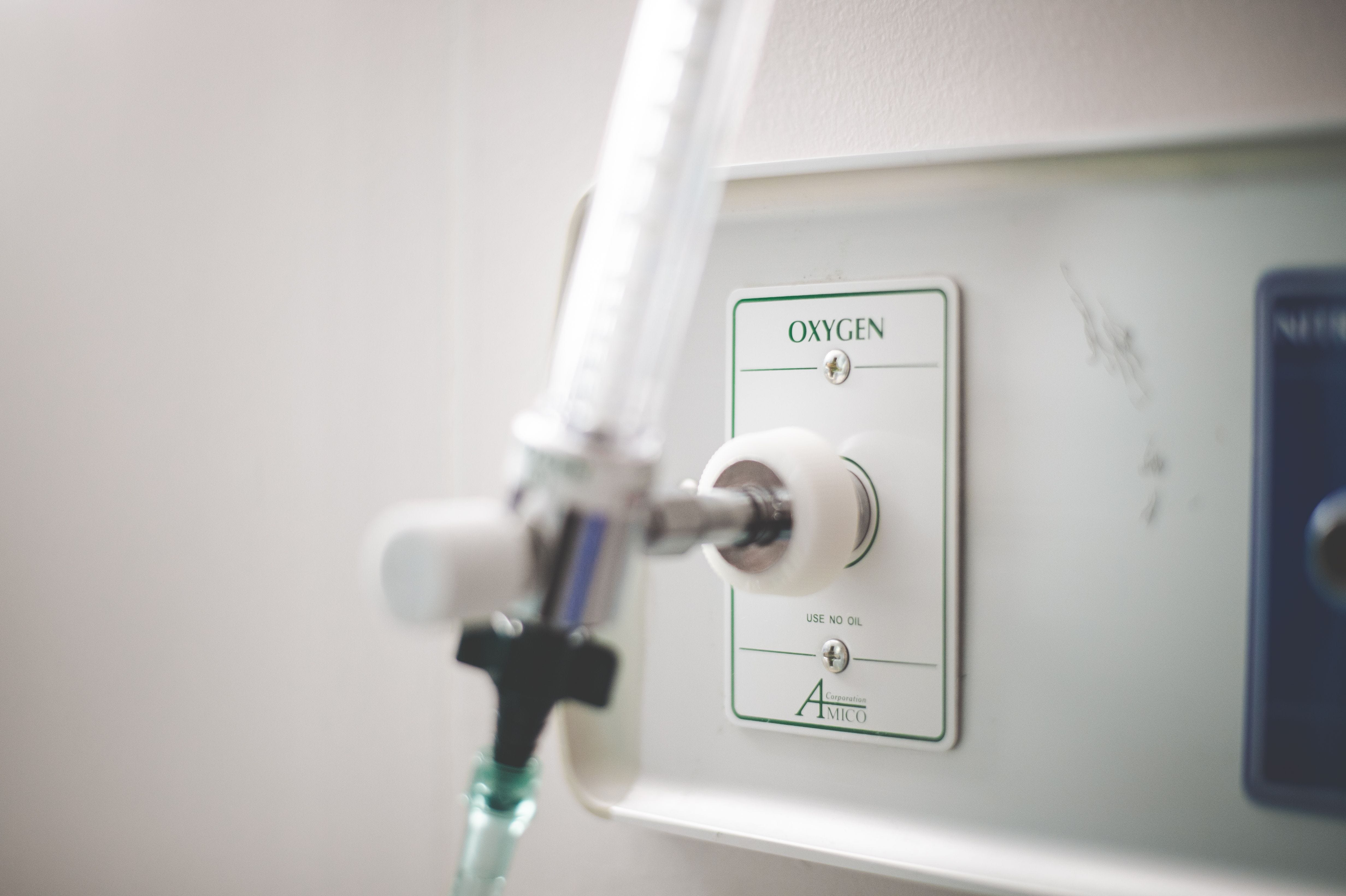
That is when his care team introduced hyperbaric oxygen therapy (HBOT), a treatment where the patient breathes 100% pure oxygen inside a pressurized chamber. The decision was made in hopes of sensitizing the tumor to ongoing treatment. Over his 5th and 6th cycles of chemoimmunotherapy, he also completed 10 sessions of HBOT per cycle. By June 2024, an abdominal CT scan showed what the care team had hoped for: all the cancer was gone, and every metastatic tumor in the peritoneum had disappeared. Tumor markers returned to normal levels, and the patient reported no new side effects. As of March 2025, the last recorded follow-up, he remained cancer-free and in good health.
This is just one case. But it reflects the potential of targeting one of cancer’s underrated hallmarks: hypoxia. Cancer hypoxia is defined as low levels of oxygen inside tumors. What does oxygen deprivation actually do to a tumor, and why does it make cancer so much harder to treat?
A Personal Detour into Existential Crisis
I first encountered the term hypoxia in a molecular cancer signaling class during my first year of graduate school. Eighty-plus slides later, I was still seeing HIF-1A eye floaters when I blinked. Signaling pathways have always fascinated me, so the information stuck. On my walks in between class and the laboratory, I would hold my breath for a set amount of time and picture all the molecular tumbles happening in my cells from my impulsive decisions (just nerdy things).
A few months later, at one of several internal research days I attended at my institution, I found out that a close colleague was studying cancer hypoxia for his dissertation. He gave a gripping oral presentation on an unprecedented method he had developed for transferring tumors between ambient air into low-oxygen environments in seconds. The method apparently involved an Olympic-style sprint (a feat only he could achieve given his height). I was pleasantly amused, as were others in the audience. But that story pulled me into his work. And it sent me spiraling into questioning my whole research hypothesis, and all research in general.
Are we doing research under the wrong conditions? If atmospheric oxygen causes molecular changes in cancer cells sitting in a plate, or in tumors removed from mice, is that a hidden variable explaining why so many drugs that work beautifully in the lab fail when they reach the clinic? I carried that question through five years of my PhD and still wonder to this day. But hey, many biological studies are not always 100% aligned with the actual human physiology.
Solid Tumors Thrive Without Air
Upon scratching the surface of this topic, I assumed cancer hypoxia is quite ironic. Picture this: cancer, a condition defined by uncontrolled growth, can thrive particularly because it is deprived of oxygen.
While the air we breathe has about 21% oxygen, normal healthy tissues maintain oxygen levels between 4.6 to 9.5% (this state is known as physoxia or normoxia). On the other hand, solid tumors often sit below 1% in a hypoxic state. As cancer cells multiply exponentially, their oxygen demand quickly outpaces the blood supply available to them. The distance between some cancer cells and blood vessels increases, preventing oxygen diffusion to tissues, and creating an even more hypoxic environment. It becomes a brutal feedback loop. And rather than dying in this oxygen-poor environment, cancer cells adapt. They rewire their metabolism and initial survival strategies, and in doing so, become harder to eliminate.
Hypoxia is present in roughly 90% of solid tumors. Cancer types that have consistently shown low oxygen levels include pancreatic cancer, head and neck cancers, breast cancer, cervical cancer, and melanoma. Studies have also linked the presence of hypoxia to lower disease-free survival in cervical cancer, prostate cancer, and head and neck squamous cell carcinoma. The more I learn about it, the clearer it becomes that hypoxia is fundamental to the progression of cancer and the success of novel therapies, and it is not talked about as often as it should.
The Protein That Senses Void
Our understanding of how cells respond to low oxygen began to crystallize in the late 1990s. In 2019, William G. Kaelin Jr., Sir Peter J. Ratcliffe, and Gregg L. Semenza were awarded the Nobel Prize in Medicine for their discovery of hypoxia-inducible factor, or HIF, the main protein that controls gene expression when oxygen levels drop.
Interest in how living things respond to oxygen deprivation actually stretches back much further. In the late 1800s, researchers were already studying hypoxia in the context of heart disease and respiratory function. Credit for recognizing hypoxia as a physiological phenomenon is often given to French physician Denis Jourdanet, who studied the effects of high altitude on human physiology. Fast forward a century later, the mechanisms that drive hypoxia are now much elucidated.
HIF exists as a two-part complex. The alpha subunit comes in three forms (HIF-1A, HIF-2A, and HIF-3A), and the beta subunit (HIF-1B) acts as its partner. HIF-1A is expressed in virtually all body tissues, while HIF-2A and HIF-3A are tissue-specific. For this article, HIF-1A is the central player.
When oxygen is present, HIF-1A is continuously tagged for destruction by a protein called von Hippel-Lindau (VHL) and degraded through a cellular disposal process called the ubiquitin pathway. So, it never gets a chance to accumulate in the cell. But when oxygen drops below the normoxic range, that destruction signal wanes. HIF-1A builds up in the cytoplasm of the cell, pairs with HIF-1B, travels into the nucleus, and begins switching on an array of genes, most of them aimed at helping the cell survive. This mechanism represents how certain cancers survive treatment assault.
A Feedback Loop Built for Survival
One of the things HIF-1A does under hypoxia is help cancer cells switch their energy source.
Normally, cells generate energy through oxidative phosphorylation, a process that requires oxygen. Under low oxygen, HIF-1A drives a shift toward glycolysis, an ancient metabolic pathway that predates the presence of oxygen in Earth’s atmosphere. Glycolysis converts glucose to pyruvate to generate energy without needing oxygen. HIF-1A enables this switch by activating genes that raise glucose uptake and glycolytic enzyme expression, like GLUT-1, HK2, and GAPDH. This metabolic flexibility makes cancer cells remarkably difficult to eliminate in a hypoxic environment. But metabolism is only an aspect of the trouble caused by hypoxia.
Oxygen distribution inside a tumor is highly uneven, fluctuating in both time and space due to unstable blood flow and ongoing blood vessel remodeling(angiogenesis). That constant variation puts pressure on cancer cells to adapt rapidly and continuously. One result of this pressure is genomic instability. Hypoxia suppresses the DNA repair mechanisms that are slow but accurate, and instead favors faster, error-prone pathways. This introduces mutations that allow cancer cells to withstand therapies designed to damage their DNA, which is exactly how many chemotherapy drugs work. So essentially, hypoxia is selecting for cancer cells that can tolerate stress and acquire aggressive traits.
The downstream consequences of unchecked HIF-1A activity eventually manifest in the clinic as increased metastasis, resistance to treatment, and reduced life expectancy.
Why the Patient’s Treatments Stopped Working
Returning to the case study, if hypoxia is this powerful, it helps explain why chemotherapy and immunotherapy had both stalled in the patient before HBOT (hyperbaric oxygen therapy) was introduced.
On the chemotherapy side, one could hypothesize this scene at the molecular level: HIF-1A and HIF-2A ramp up the production of efflux pump proteins like P-glycoprotein (P-gp) that actively pump drugs out of cells. Normally, these pumps are a defense mechanism used by healthy cells to remove toxins. Under hypoxia, cancer cells hijack the process to evade chemotherapy drugs’ effects. At the same time, as previously mentioned, hypoxia can increase the expression of DNA repair proteins, which blunts the DNA damage that chemotherapy is designed to cause. Cancer cells under low oxygen, in effect, chemotherapy-resistant. If some cancer cells were successfully eliminated with chemotherapy, the hypoxic ones wouldn’t budge. There is also a mechanical barrier created by hypoxia induced remodelling of the extracellular matrix that prevents drugs from reaching tumors effectively. So that’s another theory.
Hypoxia promotes immune system evasion in cancer. Hence, my upcoming rationale for why sintilimab did not work is also not far-fetched. The patient had received sintilimab, an immune checkpoint inhibitor that blocks a protein called PD-1, which is expressed on activated T-cells. PD-1 acts as a ‘brake’ (it dials down their T-cell’s immune activity), which helps prevent autoimmune overreaction. But then again, cancer cells and their irresistible zeal for survival exploit this by expressing a protein called PD-L1. PD-L1 binds to PD-1 to suppress the T-cell attack. So Sintilimab, which blocks PD-1 removes T-cell reluctance to attack cancer cells. Given that HIF-1A drives the expression of PD-L1 on cancer cells, the ‘brake’ on T-cells remain engaged regardless of the drug.
Beyond that, hypoxia triggers the production of cytokines that recruit regulatory T-cells (Tregs) and other immunosuppressive cells into the tumor microenvironment, thus dampening any form of immune response against cancer. So by the time immunotherapy is administered, the tumor is already surrounded by an immunosuppressive shield.
HBOT, by reoxygenating the tumor, can reverse many of these dynamics. When oxygen returns, HIF-1A degrades, efflux pumps are dialed back, DNA repair switches to its more accurate pathway(or enough reactive oxygen species are produced to cause DNA damage), angiogenesis is impaired, and the immunosuppressive environment starts to dismantle. That is likely why adding HBOT to the patient’s existing regimen produced such a dramatic response when the regimen alone had not.
How Do We Detect and Target Hypoxia?
Given how consequential hypoxia is, detecting it matters. In clinical settings, hypoxic regions of tumors can be visualized using non-invasive techniques like Positron Emission Tomography (PET) tracers. MRI is also used in some cases, though concerns remain about its sensitivity and resolution.
When hypoxia is suspected to be driving resistance, multiple approaches can be taken to sensitize tumors to treatment. One approach, of course, is to improve oxygen levels within tumors, such as through HBOT, carbogen inhalation (95% oxygen + 5% carbon dioxide), or nanomaterials that release oxygen precisely to hypoxic tumors. Although only established as a standard of care in Scandinavia, nimorazole, an oxygen-mimetic radiosensitizer, is used with radiotherapy for head and neck squamous cell carcinoma based on the DAHANCA-5 trial showing improved tumor control.
Another strategy is to inhibit HIF and prevent its accumulation in cells. Small-molecule HIF inhibitors such as camptothecin and PX-478 have reached clinical trials. Researchers also use hypoxia-activated prodrugs (HAPs), drugs that remain inert under normal oxygen conditions but activate specifically in the oxygen-poor regions of a tumor. One example of HAP success is tirapazamine (TPZ), which showed benefit when combined with cisplatin and radiotherapy in a phase II head and neck cancer trial.
The mTOR inhibitor everolimus, tested in phase III clinical trials, prolonged progression-free survival in patients with advanced renal cell carcinoma. The literature suggests that this benefit may be partly due to reduced HIF-1α activity. Hypoxic tumors also have a low pH, so nanoparticles designed to target acidic environments could effectively and precisely target hypoxic regions.
Hypoxia is a rich target because of how deeply embedded it is in tumor biology. Some clinical studies have demonstrated that targeting hypoxia as a supplementary cancer treatment can provide significant clinical benefit and enhance treatment efficacy. Also, one case study reported that in a colorectal cancer patient, hypoxia was found to explain the sudden aggressive course of the tumor. Similarly, in two patients with metastatic colorectal cancer not responding to chemotherapy, a hypoxia-activated epigenetic drug helped resensitize their tumors to treatment. Hypoxia is also being used as a biomarker of distant metastasis, and thus guiding patient selection for certain treatments. These examples suggest that detecting and managing hypoxia could change diagnosis, prognosis, and treatment decisions for individual patients.
Where This Leaves Us
After going down the rabbit hole of literature studies and clinical trials, there is no doubt that hypoxia is a major driver of cancer treatment resistance. It is also a known enabler of metastasis, genomic instability, immune evasion, and metabolic adaptation. In short, it aids and abets nearly every feature that makes cancer deadly.
This very involvement makes it a compelling target. The more we understand the mechanisms HIF-1A triggers, the more potential intervention points emerge. Success in addressing hypoxia will depend on better detection tools, smarter patient stratification, and treatment combinations specifically designed with the hypoxic tumor microenvironment in mind.
The questions I had during my existential crisis are now layered. Beyond the notion that we may have been approaching cancer research the wrong way for decades, should targeting hypoxia become standard in cases of chemotherapeutic resistance? Should tumor hypoxia always be measured during treatment and monitoring to guide clinical decisions? I am no oncologist. But that one case report of the 62-year-old patient did exemplify how resistance can be overcome when hypoxia is identified as the culprit. So I wonder how many patients have been classified as “non-responders” when they actually might respond given the right conditions?
References
1. West JB, Richalet JP. Denis Jourdanet (1815-1892) and the early recognition of the role of hypoxia at high altitude. Am J Physiol Lung Cell Mol Physiol. 2013;305(5):L333-L340. doi:10.1152/ajplung.00128.2013
2. Hompland T, Fjeldbo CS, Lyng H. Tumor Hypoxia as a Barrier in Cancer Therapy: Why Levels Matter. Cancers. 2021; 13(3):499. https://doi.org/10.3390/cancers13030499
3. Lee CC, Wu CY, Yang HY. Discoveries of how cells sense oxygen win the 2019 Nobel Prize in Physiology or medicine. Biomed J. 2020;43(5):434-437. doi:10.1016/j.bj.2020.05.019
4. Li W, Wei J, Zhang P, et al. Hyperbaric oxygen therapy as an immunosensitizing strategy in advanced gastric hepatoid adenocarcinoma: a case report. Front Immunol. 2025;16:1625273. Published 2025 Jun 27. doi:10.3389/fimmu.2025.1625273
5. Kunachowicz D, Tomecka P, Sędzik M, Kalinin J, Kuźnicki J, Rembiałkowska N. Influence of Hypoxia on Tumor Heterogeneity, DNA Repair, and Cancer Therapy: From Molecular Insights to Therapeutic Strategies. Cells. 2025;14(14):1057. Published 2025 Jul 10. doi:10.3390/cells14141057
6. Chen Z, Han F, Du Y, Shi H, Zhou W. Hypoxic microenvironment in cancer: molecular mechanisms and therapeutic interventions. Signal Transduct Target Ther. 2023;8(1):70. Published 2023 Feb 17. doi:10.1038/s41392-023-01332-8
7. Fleming IN, Manavaki R, Blower PJ, et al. Imaging tumour hypoxia with positron emission tomography. Br J Cancer. 2015;112(2):238-250. doi:10.1038/bjc.2014.610
8. Reid T, Dad S, Korn R, Oronsky B, Knox S, Scicinski J. Two Case Reports of Resensitization to Previous Chemotherapy with the Novel Hypoxia-Activated Hypomethylating Anticancer Agent RRx-001 in Metastatic Colorectal Cancer Patients. Case Rep Oncol. 2014;7(1):79-85. Published 2014 Jan 24. doi:10.1159/000358382
9. Muz B, de la Puente P, Azab F, Azab AK. The role of hypoxia in cancer progression, angiogenesis, metastasis, and resistance to therapy. Hypoxia (Auckl). 2015;3:83-92. Published 2015 Dec 11. doi:10.2147/HP.S93413
10. Laws K, Murray GI, Kerr K, et al. Hypoxia PET/CT and colorectal cancer: a case report. August 2020. https://hdl.handle.net/2134/12813962.v1. Accessed May 29, 2026.
11. Gui C, Wray R, Schöder H, et al. Tumor Hypoxia on 18F-fluoromisonidazole Positron Emission Tomography and Distant Metastasis From Head and Neck Squamous Cell Carcinoma. JAMA Netw Open. 2024;7(9):e2436407. Published 2024 Sep 3. doi:10.1001/jamanetworkopen.2024.36407
12. Bigos KJ, Quiles CG, Lunj S, et al. Tumour response to hypoxia: understanding the hypoxic tumour microenvironment to improve treatment outcome in solid tumours. Front Oncol. 2024;14:1331355. Published 2024 Jan 30. doi:10.3389/fonc.2024.1331355
13. Więch-Walów A, Bartoszewska S, Barton A, et al. HIF inhibition: Current strategies and clinical challenges. Redox Biol. 2026;94:104220. doi:10.1016/j.redox.2026.104220
Really interesting topic and it’s not infrequent I have patients asking me about adding hyperbaric oxygen to standard of care treatments. Given a low risk intervention I think it’s worth exploring bringing clinical trials to community cancer centers that already have access to HBOT. At the very least it’s worth more discussion among oncology researchers. I’ll look for anything out there at ASCO this weekend. The only limitation I see in that case is that delayed responses are not uncommon with immunotherapy sometimes 4 or 5 months after starting treatment we start to see responses in immunogenic cancers so that may have been an explanation of response in that case.